Figure 9
CT signs of pressure induced expansion of paranasal sinus structures
Peter AR Clement* and Stijn Halewyck
Published: 26 September, 2017 | Volume 1 - Issue 1 | Pages: 077-087
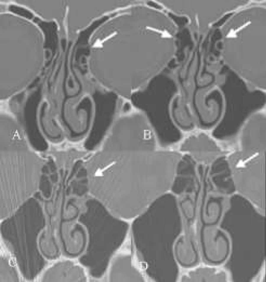
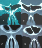
Figure 9:
(A-D) Coronal CT-scans. Some rounding of supra-orbital recess (A), hyperinflated ethmoid (with ballooning of both lamina orbitalis, B,C) with clear-cut “cobble-stone”aspect of this orbital lamina on the left side (B,C), hyperinflated bullae bilaterally (B,C) septal deviation to the right and beginning disease of the mucosa in and around in the infundibulum ethmoidale of the left side (A,B,C), and a left concha media bullosa (A).
Read Full Article HTML DOI: 10.29328/journal.hor.1001013 Cite this Article Read Full Article PDF
More Images
Similar Articles
-
CT signs of pressure induced expansion of paranasal sinus structuresPeter AR Clement*,Stijn Halewyck. CT signs of pressure induced expansion of paranasal sinus structures. . 2017 doi: 10.29328/journal.hor.1001013; 1: 077-087
Recently Viewed
-
Pharmacovigilance is Important for Assessments of Drugs, and Withdrawal of the Drugs that have Adverse Effects More than The Benefits of Their TreatmentRezk R Ayyad,Yasser Abdel Allem Hassan,Ahmed R Ayyad*. Pharmacovigilance is Important for Assessments of Drugs, and Withdrawal of the Drugs that have Adverse Effects More than The Benefits of Their Treatment. Arch Pharm Pharma Sci. 2025: doi: 10.29328/journal.apps.1001069; 9: 042-045
-
Ectopic Pregnancy Risk Factors Presentation and Management OutcomesAwadalla Abdelwahid Suliman*, Hajar Suliman Ibrahim Ahmed, Kabbashi Mohammed Adam Hammad, Ibtehal Jaffer Youssef Alsiddig, Mohamed Abdalla Elamin Abdelgader, Abdallah Omer Elzein Elhag, Safa Mohamed Ibrahim. Ectopic Pregnancy Risk Factors Presentation and Management Outcomes. Clin J Obstet Gynecol. 2023: doi: 10.29328/journal.cjog.1001143; 6: 143-149
-
Role of Perianesthesia Nurses in Enhanced Recovery After Surgery (ERAS) Protocols: A Narrative Review and Comparative Outcomes AnalysisOghogho Linda Akarogbe*,Geneva Igwama,Olachi Lovina Emenyonu,Idowu M Ariyibi. Role of Perianesthesia Nurses in Enhanced Recovery After Surgery (ERAS) Protocols: A Narrative Review and Comparative Outcomes Analysis. Int J Clin Anesth Res. 2025: doi: 10.29328/journal.ijcar.1001034; 9: 037-039
-
Assessment of Albino Beech Supremacy to Pigmented Beech Proves to Be A Better Environmental Condition BioindicatorRenata Gagić-Serdar*,Miroslava Marković,Ljubinko Rakonjac,Goran Češljar,Bojan Konatar. Assessment of Albino Beech Supremacy to Pigmented Beech Proves to Be A Better Environmental Condition Bioindicator. Insights Biol Med. 2025: doi: 10.29328/journal.ibm.1001031; 9: 009-015.
-
Minds after Death: The Expanding Role of Psychological Autopsy in Investigations: A ReviewIshan Jain*,Oindrila Mahapatra,Yogesh Kumar. Minds after Death: The Expanding Role of Psychological Autopsy in Investigations: A Review. J Forensic Sci Res. 2025: doi: 10.29328/journal.jfsr.1001096; 9: 155-0
Most Viewed
-
Feasibility study of magnetic sensing for detecting single-neuron action potentialsDenis Tonini,Kai Wu,Renata Saha,Jian-Ping Wang*. Feasibility study of magnetic sensing for detecting single-neuron action potentials. Ann Biomed Sci Eng. 2022 doi: 10.29328/journal.abse.1001018; 6: 019-029
-
Evaluation of In vitro and Ex vivo Models for Studying the Effectiveness of Vaginal Drug Systems in Controlling Microbe Infections: A Systematic ReviewMohammad Hossein Karami*, Majid Abdouss*, Mandana Karami. Evaluation of In vitro and Ex vivo Models for Studying the Effectiveness of Vaginal Drug Systems in Controlling Microbe Infections: A Systematic Review. Clin J Obstet Gynecol. 2023 doi: 10.29328/journal.cjog.1001151; 6: 201-215
-
Prospective Coronavirus Liver Effects: Available KnowledgeAvishek Mandal*. Prospective Coronavirus Liver Effects: Available Knowledge. Ann Clin Gastroenterol Hepatol. 2023 doi: 10.29328/journal.acgh.1001039; 7: 001-010
-
Causal Link between Human Blood Metabolites and Asthma: An Investigation Using Mendelian RandomizationYong-Qing Zhu, Xiao-Yan Meng, Jing-Hua Yang*. Causal Link between Human Blood Metabolites and Asthma: An Investigation Using Mendelian Randomization. Arch Asthma Allergy Immunol. 2023 doi: 10.29328/journal.aaai.1001032; 7: 012-022
-
An algorithm to safely manage oral food challenge in an office-based setting for children with multiple food allergiesNathalie Cottel,Aïcha Dieme,Véronique Orcel,Yannick Chantran,Mélisande Bourgoin-Heck,Jocelyne Just. An algorithm to safely manage oral food challenge in an office-based setting for children with multiple food allergies. Arch Asthma Allergy Immunol. 2021 doi: 10.29328/journal.aaai.1001027; 5: 030-037

HSPI: We're glad you're here. Please click "create a new Query" if you are a new visitor to our website and need further information from us.
If you are already a member of our network and need to keep track of any developments regarding a question you have already submitted, click "take me to my Query."


