Figure 2
CT signs of pressure induced expansion of paranasal sinus structures
Peter AR Clement* and Stijn Halewyck
Published: 26 September, 2017 | Volume 1 - Issue 1 | Pages: 077-087
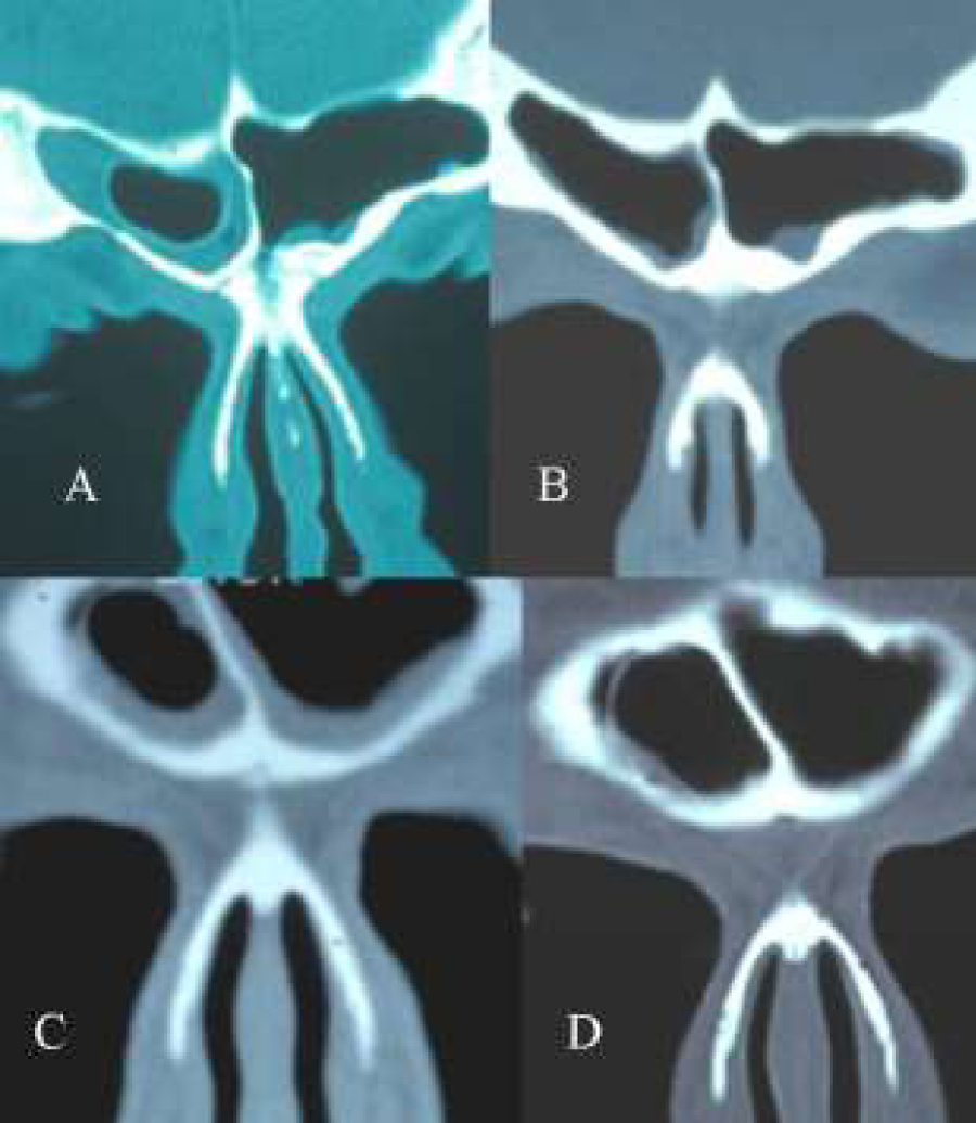
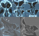
Figure 2:
(A-D) Coronal CT-scans. A+B: pre-and postoperative CT-scan of a frontal cell invading the frontal sinus. The interior of the cell is free of disease. After removal of the cell the mucosa of the frontal sinus becomes normal. C+D: Two CT-scans at the same level with a 2 year difference in a patient with a frontal cell in the frontal sinus. Note that the cell has increased in size (D).
Read Full Article HTML DOI: 10.29328/journal.hor.1001013 Cite this Article Read Full Article PDF
More Images
Similar Articles
-
CT signs of pressure induced expansion of paranasal sinus structuresPeter AR Clement*,Stijn Halewyck. CT signs of pressure induced expansion of paranasal sinus structures. . 2017 doi: 10.29328/journal.hor.1001013; 1: 077-087
Recently Viewed
-
Harmonizing Artificial Intelligence Governance; A Model for Regulating a High-risk Categories and Applications in Clinical Pathology: The Evidence and some ConcernsMaxwell Omabe*. Harmonizing Artificial Intelligence Governance; A Model for Regulating a High-risk Categories and Applications in Clinical Pathology: The Evidence and some Concerns. Arch Pathol Clin Res. 2024: doi: 10.29328/journal.apcr.1001040; 8: 001-005
-
The Accuracy of pHH3 in Meningioma Grading: A Single Institution StudyMansouri Nada1, Yaiche Rahma*, Takout Khouloud, Gargouri Faten, Tlili Karima, Rachdi Mohamed Amine, Ammar Hichem, Yedeas Dahmani, Radhouane Khaled, Chkili Ridha, Msakni Issam, Laabidi Besma. The Accuracy of pHH3 in Meningioma Grading: A Single Institution Study. Arch Pathol Clin Res. 2024: doi: 10.29328/journal.apcr.1001041; 8: 006-011
-
Autoantibodies in Autoimmune Addison’s Disease: Why are they Important?Maria Rosaria De Cagna, Norma Notaristefano, Maurizio Schiavone, Gianluca Palatella, Federica Ranù, Carmela Presicci, Valerio Cecinati, Marilina Tampoia*. Autoantibodies in Autoimmune Addison’s Disease: Why are they Important?. Arch Pathol Clin Res. 2024: doi: 10.29328/journal.apcr.1001042; 8: 012-015
-
A Rare Case of Uvulitis Following Endotracheal Intubation Using C- Mac Videolaryngoscope and How To Avoid ItKiran Kuruvilla George, Jini Chirackel Thomas, Krishnan B Sivaram, Krishna Prasad T*. A Rare Case of Uvulitis Following Endotracheal Intubation Using C- Mac Videolaryngoscope and How To Avoid It. Arch Pathol Clin Res. 2024: doi: 10.29328/journal.apcr.1001043; 8: 016-020
-
Immune-mediated neuropathy related to bortezomib in a patient with multiple myelomaSusanne Koeppen*,Jörg Hense,Kay Wilhelm Nolte,Joachim Weis. Immune-mediated neuropathy related to bortezomib in a patient with multiple myeloma. Arch Pathol Clin Res. 2022: doi: 10.29328/journal.apcr.1001028; 6: 001-004
Most Viewed
-
Feasibility study of magnetic sensing for detecting single-neuron action potentialsDenis Tonini,Kai Wu,Renata Saha,Jian-Ping Wang*. Feasibility study of magnetic sensing for detecting single-neuron action potentials. Ann Biomed Sci Eng. 2022 doi: 10.29328/journal.abse.1001018; 6: 019-029
-
Evaluation of In vitro and Ex vivo Models for Studying the Effectiveness of Vaginal Drug Systems in Controlling Microbe Infections: A Systematic ReviewMohammad Hossein Karami*, Majid Abdouss*, Mandana Karami. Evaluation of In vitro and Ex vivo Models for Studying the Effectiveness of Vaginal Drug Systems in Controlling Microbe Infections: A Systematic Review. Clin J Obstet Gynecol. 2023 doi: 10.29328/journal.cjog.1001151; 6: 201-215
-
Prospective Coronavirus Liver Effects: Available KnowledgeAvishek Mandal*. Prospective Coronavirus Liver Effects: Available Knowledge. Ann Clin Gastroenterol Hepatol. 2023 doi: 10.29328/journal.acgh.1001039; 7: 001-010
-
Causal Link between Human Blood Metabolites and Asthma: An Investigation Using Mendelian RandomizationYong-Qing Zhu, Xiao-Yan Meng, Jing-Hua Yang*. Causal Link between Human Blood Metabolites and Asthma: An Investigation Using Mendelian Randomization. Arch Asthma Allergy Immunol. 2023 doi: 10.29328/journal.aaai.1001032; 7: 012-022
-
An algorithm to safely manage oral food challenge in an office-based setting for children with multiple food allergiesNathalie Cottel,Aïcha Dieme,Véronique Orcel,Yannick Chantran,Mélisande Bourgoin-Heck,Jocelyne Just. An algorithm to safely manage oral food challenge in an office-based setting for children with multiple food allergies. Arch Asthma Allergy Immunol. 2021 doi: 10.29328/journal.aaai.1001027; 5: 030-037

HSPI: We're glad you're here. Please click "create a new Query" if you are a new visitor to our website and need further information from us.
If you are already a member of our network and need to keep track of any developments regarding a question you have already submitted, click "take me to my Query."


