Figure 1
Recurrent Mucoepidermoid Carcinoma of Parotid with Facial Tics - Report of an unusual case
Pirabu Sakthivel*, Chirom Amit Singh, Smriti Panda, Suresh Chandra Sharma, Konki Malla Abhilash and Milind Sagar
Published: 16 June, 2017 | Volume 1 - Issue 1 | Pages: 032-036
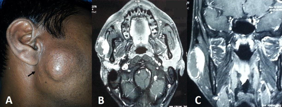
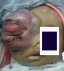
Figure 1:
A Clinical picture showing preauricular swelling and scar of previous surgery (arrow). B&C. Axial and coronal MRI post contrast T1 weighted images show peripheral enhancement of the cystic component and intense enhancement of the solid component.
Read Full Article HTML DOI: 10.29328/journal.hor.1001006 Cite this Article Read Full Article PDF
More Images
Similar Articles
-
Recurrent Mucoepidermoid Carcinoma of Parotid with Facial Tics - Report of an unusual casePirabu Sakthivel*,Chirom Amit Singh,Smriti Panda,Suresh Chandra Sharma,Konki Malla Abhilash,Milind Sagar. Recurrent Mucoepidermoid Carcinoma of Parotid with Facial Tics - Report of an unusual case. . 2017 doi: 10.29328/journal.hor.1001006; 1: 032-036
Recently Viewed
-
Effectiveness of an Ayurvedic Gel for Tooth Pain Relief Due to Dental Caries: A Randomized Controlled TrialNandlal Bhojraj*, Prem K Sreenivasan, Paras Mull Gehlot, Vinutha Manjunath, Manjunath MK. Effectiveness of an Ayurvedic Gel for Tooth Pain Relief Due to Dental Caries: A Randomized Controlled Trial. J Clin Adv Dent. 2024: doi: 10.29328/journal.jcad.1001041; 8: 013-019
-
Investigating the Effect of the Family-Centered Empowerment Model (FCEM) on the Empowerment Indicators of Student Girls with Iron Deficiency Anemia (IDA) and Their MothersFatemeh Alhani,Hasan Navipor,Fatemeh Sadat Seyed Nematollah Roshan*. Investigating the Effect of the Family-Centered Empowerment Model (FCEM) on the Empowerment Indicators of Student Girls with Iron Deficiency Anemia (IDA) and Their Mothers. Insights Depress Anxiety. 2025: doi: 10.29328/journal.ida.1001045; 9: 017-024
-
Novel Mutation in Famous Gene Diseases in Red Blood CellsMahdi Nowroozi*. Novel Mutation in Famous Gene Diseases in Red Blood Cells. New Insights Obes Gene Beyond. 2025: doi: 10.29328/journal.niogb.1001023; 9: 013-020
-
Acute Gas Toxicity at Work: A Tale of Two Cases with Review of LiteratureRishabh Kumar Singh,Jitender Pratap Singh,Manjari Kishore*,HM Garg. Acute Gas Toxicity at Work: A Tale of Two Cases with Review of Literature. J Forensic Sci Res. 2025: doi: 10.29328/journal.jfsr.1001091; 9: 125-128
-
Minds after Death: The Expanding Role of Psychological Autopsy in Investigations: A ReviewIshan Jain*,Oindrila Mahapatra,Yogesh Kumar. Minds after Death: The Expanding Role of Psychological Autopsy in Investigations: A Review. J Forensic Sci Res. 2025: doi: 10.29328/journal.jfsr.1001096; 9: 155-0
Most Viewed
-
Feasibility study of magnetic sensing for detecting single-neuron action potentialsDenis Tonini,Kai Wu,Renata Saha,Jian-Ping Wang*. Feasibility study of magnetic sensing for detecting single-neuron action potentials. Ann Biomed Sci Eng. 2022 doi: 10.29328/journal.abse.1001018; 6: 019-029
-
Evaluation of In vitro and Ex vivo Models for Studying the Effectiveness of Vaginal Drug Systems in Controlling Microbe Infections: A Systematic ReviewMohammad Hossein Karami*, Majid Abdouss*, Mandana Karami. Evaluation of In vitro and Ex vivo Models for Studying the Effectiveness of Vaginal Drug Systems in Controlling Microbe Infections: A Systematic Review. Clin J Obstet Gynecol. 2023 doi: 10.29328/journal.cjog.1001151; 6: 201-215
-
Prospective Coronavirus Liver Effects: Available KnowledgeAvishek Mandal*. Prospective Coronavirus Liver Effects: Available Knowledge. Ann Clin Gastroenterol Hepatol. 2023 doi: 10.29328/journal.acgh.1001039; 7: 001-010
-
Causal Link between Human Blood Metabolites and Asthma: An Investigation Using Mendelian RandomizationYong-Qing Zhu, Xiao-Yan Meng, Jing-Hua Yang*. Causal Link between Human Blood Metabolites and Asthma: An Investigation Using Mendelian Randomization. Arch Asthma Allergy Immunol. 2023 doi: 10.29328/journal.aaai.1001032; 7: 012-022
-
An algorithm to safely manage oral food challenge in an office-based setting for children with multiple food allergiesNathalie Cottel,Aïcha Dieme,Véronique Orcel,Yannick Chantran,Mélisande Bourgoin-Heck,Jocelyne Just. An algorithm to safely manage oral food challenge in an office-based setting for children with multiple food allergies. Arch Asthma Allergy Immunol. 2021 doi: 10.29328/journal.aaai.1001027; 5: 030-037

HSPI: We're glad you're here. Please click "create a new Query" if you are a new visitor to our website and need further information from us.
If you are already a member of our network and need to keep track of any developments regarding a question you have already submitted, click "take me to my Query."


